
Most people associate good eyesight with being able to read small text or see objects clearly in the distance. However, sharp central vision is only one part of how we see and interact with the world around us.
Your visual field includes everything you can see while looking straight ahead. This covers the detailed central area you use for reading and recognising faces, as well as the wider peripheral vision that helps you notice movement, navigate busy spaces and remain aware of objects approaching from the side.
A visual field test assesses how sensitive different areas of your vision are. It may be recommended when an optometrist wants to investigate possible changes involving the retina, optic nerve or visual pathways connecting the eyes and brain.
Goldsmith & Harvey’s experienced Bristol opticians provide professional examinations and personalised advice for patients who want to understand and protect their long-term eye health.
What Is a Visual Field Test?
A visual field test measures how much you can see around you while your eyes remain focused on a central point. It checks both central vision and peripheral, or side, vision.
During an automated test, small lights normally appear in different positions within a bowl-shaped machine. Some lights are relatively bright, while others may be very faint. You press a button whenever you see one, allowing the equipment to map how sensitive different parts of your vision are.
Each eye is usually tested separately. This is important because the visual fields of both eyes overlap during normal daily activities. Testing them individually can reveal a change in one eye that may not be noticeable when both eyes are open.
Guy’s and St Thomas’ NHS Foundation Trust explains that visual field testing can assess areas of vision associated with the retina, optic nerve and visual pathway through the brain.
What Is the Difference Between Central and Peripheral Vision?
Central vision is the detailed sight produced by the area you are looking directly towards. It allows you to read, examine fine details, recognise faces and complete tasks that require precision.
Peripheral vision is everything you see outside this direct central area. Although objects in your peripheral vision may not appear as sharply detailed, this wider awareness is essential for detecting movement and understanding what is happening around you.
You use peripheral vision when walking through a crowded shop, crossing a road, playing sport or noticing somebody approaching from the side. It also contributes to spatial awareness and your ability to navigate unfamiliar environments.
A person can retain clear central vision while experiencing reduced sensitivity in parts of their peripheral vision. This is why reading an eye chart successfully does not always provide a complete assessment of how well somebody sees across their entire visual field.
Why Can Visual Field Loss Be Difficult to Notice?
Changes to peripheral vision may develop gradually. When this happens, the brain can adapt to the missing information, making the reduction difficult to recognise during everyday activities.
The visual fields of both eyes also overlap considerably. If one eye develops an area of reduced sensitivity, the other may partly compensate while both eyes remain open.
Somebody with early field loss may therefore continue to read clearly, watch television and complete normal close-up tasks. They may not experience the type of blur commonly associated with needing a new glasses prescription.
This does not mean that visual field loss has no impact. More advanced changes may affect mobility, driving, awareness of obstacles or the ability to notice hazards approaching from the side. University Hospitals Sussex notes that mild field loss may go unnoticed when central vision remains clear and changes develop slowly.
What Happens During a Visual Field Test?
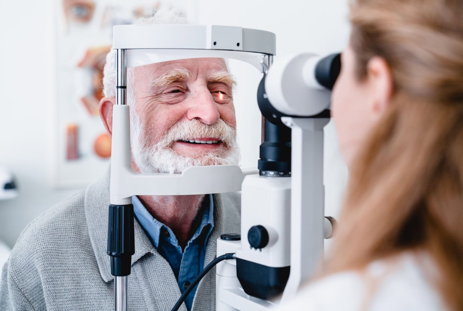
During a commonly used automated visual field test, you sit in front of the machine with your chin and forehead resting against supports. One eye is tested at a time while the other is covered.
You will be asked to look continuously at a fixed point in the centre of the machine. Lights then appear in different positions and at different brightness levels. Each time you see a light, you press a handheld button.
It is important to keep looking at the central target rather than moving your eyes towards each flash. The test is designed to measure what you can detect outside the area you are looking at directly. You will not see every light, and there may be periods when nothing appears to happen. This is a normal part of the test. The equipment is trying to determine the dimmest light you can detect in different areas of your vision.
Is a Visual Field Test Difficult?
Visual field testing is not painful, but it does require concentration. Some patients find it unusual because they must keep looking straight ahead while responding to lights that appear elsewhere.
You may worry that you have missed a light or pressed the button at the wrong time. Occasional missed or accidental responses are expected, and the equipment includes checks that help the clinician assess how reliable the result is.
Try to remain relaxed, blink normally and respond only when you believe you have seen a light. There is no benefit in guessing. If you become tired, uncomfortable or uncertain about the instructions, tell the person carrying out the assessment.
The test can often be paused if you need a short break. Good positioning and concentration help produce more reliable results, but patients are not expected to perform perfectly.
How Long Does a Visual Field Test Take?
The length of a visual field test varies according to the equipment, the area being assessed and how quickly you respond.
An automated Humphrey visual field test commonly used for glaucoma assessment may take approximately five to ten minutes for each eye. Other forms of testing can be shorter or considerably longer.
The complete appointment may also include checks of your vision, eye pressure, prescription and internal eye health. The optometrist may need to discuss your symptoms and medical history or compare results with previous examinations.
It is important not to rush the process. A steady position and consistent concentration can improve the reliability of the test, even when this means taking a short break between eyes.
Why Might You Need a Visual Field Test?
An optometrist may recommend visual field testing when there is a reason to examine your peripheral vision more closely. This recommendation could follow a routine eye examination, a change in symptoms or a finding involving the optic nerve.
The test is frequently associated with glaucoma because the condition can damage the optic nerve and affect peripheral vision. It may also be used when glaucoma is suspected because of raised eye pressure, the appearance of the optic nerve or a relevant family history.
Visual field assessment can also provide useful information when investigating conditions involving the retina or the visual pathways within the brain. In hospital settings, it may be used as part of neurological assessments or following events such as a stroke.
A visual field test does not usually provide a complete diagnosis on its own. Its results must be considered alongside the wider eye examination and any other appropriate investigations.
How Is Visual Field Testing Used for Glaucoma?
Glaucoma involves damage to the optic nerve, which carries information from the eye to the brain. In many cases, peripheral vision can be affected before central sight begins to change.
Visual field testing helps identify patterns of reduced sensitivity that may be associated with optic nerve damage. If a patient has already been diagnosed with glaucoma, repeating the test can help monitor whether their field of vision is stable or changing.
The result is normally considered alongside other information. This may include measurements of eye pressure, the appearance of the optic nerve, corneal thickness, OCT images and the patient’s family and medical history.
An unusual visual field result does not automatically mean that somebody has glaucoma. Fatigue, concentration, cataracts, eyelid position and other factors may influence the result. Further testing or repeated assessment may be required. Patients can arrange an assessment through Goldsmith & Harvey’s glaucoma treatment in Bristol service.
Can You Have Field Loss With Good Central Vision?
It is possible to have clear central vision while experiencing reduced peripheral vision. This is because the detailed vision measured by an eye chart only assesses a small part of your overall visual field.
Somebody may be able to read the smallest line of letters and still have areas where their side vision is less sensitive. The change may not be immediately obvious, particularly when it develops slowly or affects one eye more than the other.
This distinction is one reason a comprehensive eye examination may include several different tests. Visual acuity, prescription checks, eye pressure measurements, retinal examination, OCT imaging and visual field testing all assess different aspects of vision and eye health.
No single result tells the whole story. Your optometrist selects the most appropriate combination of tests according to your age, symptoms, eye health and personal risk factors.
Can a Visual Field Test Detect Neurological Problems?
Vision does not depend on the eyes alone. Information captured by the retina travels through the optic nerves and along visual pathways within the brain before it is processed into the images we understand.
A problem affecting any part of this pathway may produce a particular pattern of visual field loss. Visual field tests can therefore provide information that supports the investigation of certain neurological conditions.
This does not mean that the test can independently diagnose a stroke, brain condition or neurological disorder. If the pattern raises concern, the patient may require further assessment, which could include examination by an ophthalmologist, neurologist or another appropriate medical professional.
Sudden vision loss, new double vision, severe eye pain or sudden changes affecting part of your visual field should not be left until a routine appointment. These symptoms require prompt professional assessment. Guy’s and St Thomas’ states that visual field tests are regularly used in both glaucoma and neurology clinics.
Why Might the Test Need to Be Repeated?
Being asked to repeat a visual field test does not necessarily mean that a serious problem has been found. The assessment requires concentration, and first-time patients may need an opportunity to become familiar with the process.
A result may be less reliable if you look away from the central target, become tired, press the button when no light is present or struggle to maintain a comfortable position. Dry or watery eyes may also make concentration more difficult.
Repeating the test can help determine whether an unusual area appears consistently or was caused by the testing process. When the same pattern is reproduced across several reliable tests, the finding may carry greater clinical significance.
For patients with a diagnosed condition, repeated testing creates a record that can be compared over time. This allows eye-care professionals to assess whether the visual field remains stable or shows evidence of progression.
What Do Visual Field Test Results Look Like?
Visual field results are commonly displayed as maps or charts showing how sensitive different areas of each eye are. The clinician can use these maps to identify areas where lights were not seen at the expected brightness.
The result may be compared with typical sensitivity levels for people of a similar age. This is important because visual sensitivity naturally changes over time, and the interpretation should account for the patient’s age and wider clinical circumstances.
The equipment also produces reliability measurements. These help the clinician understand whether the patient maintained focus, responded consistently and completed the test in a way that makes the result suitable for interpretation.
Patients should not attempt to diagnose themselves by looking at the shaded areas or numerical values on a printout. The pattern must be interpreted alongside the eye examination, symptoms, previous results and other clinical tests.
Does an Abnormal Result Always Mean Eye Disease?
An unusual visual field result does not automatically confirm an eye condition. Several factors can affect the quality or appearance of the test.
A glasses frame may obstruct part of the peripheral view, which is why a special lens may be placed within the machine. A drooping eyelid, cataract, tiredness or difficulty following the instructions may also influence the result.
The location and pattern of any field change are important. Different conditions can produce different patterns, but further information is normally needed before a diagnosis can be reached.
Your optometrist may recommend repeating the visual field test, examining the optic nerve, measuring eye pressure, completing an OCT scan or referring you for specialist assessment. University Hospitals Sussex emphasises that visual field testing is only one part of the overall assessment required to establish why field loss is present.
How Should You Prepare for a Visual Field Test?
Bring the glasses you normally use, including any reading glasses, to your appointment. The person performing the test will decide whether you should wear your own correction or use a lens built into the equipment.
Try to attend when you are able to concentrate and follow the instructions comfortably. You do not need to practise beforehand or worry about responding to every light.
During the test, remain focused on the central target, blink normally and press the button only when you see a light. Tell the clinician if the target becomes blurry, your eye feels uncomfortable or you need the instructions repeated.
Most importantly, do not treat the assessment as an examination you can pass or fail. The purpose is to record an accurate representation of your vision, not to achieve a particular score.
How Often Will You Need Visual Field Testing?
The frequency of visual field testing depends on why it was recommended. Somebody undergoing an initial investigation may be asked to repeat the test relatively soon so that the results can be confirmed.
Patients with glaucoma or another condition affecting the visual field may require ongoing assessments. The interval between tests will be based on their diagnosis, level of risk, previous results and the advice of the professional managing their care.
Some patients may be tested annually, while others require more frequent monitoring. There is no single timetable that is suitable for everyone.
Following the recommended schedule is important because comparisons between reliable tests help identify whether the field of vision remains stable. You should not delay a review simply because your central vision still appears clear.
Regular adult eye tests in Bristol provide an opportunity to review your eye health and decide whether additional assessment is appropriate.
Arrange a Professional Eye Examination in Bristol
A visual field test provides information that cannot be obtained from a standard letter chart alone. By mapping both central and peripheral vision, it can support the assessment of glaucoma, optic nerve changes, retinal conditions and concerns involving the visual pathways.
The result is most useful when interpreted as part of a wider eye-health assessment. Your symptoms, prescription, eye pressure, optic nerve appearance, medical history and previous test results may all influence the advice you receive.
Goldsmith & Harvey provides professional eye examinations from its convenient practice inside the Asda Longwell Green Supercentre. Patients who may qualify for funded examinations can also learn more about the practice’s NHS eye tests in Bristol.
Book an appointment with Goldsmith & Harvey today if you have noticed a change in your vision, have concerns about your peripheral sight or have been advised to arrange further eye-health testing.